| |
For most of us, Valentine's Day conjures images of roses, chocolate, or a candlelit dinner for two. In place of such traditional Valentine fare, attendees of this year's Biophysical Society meeting were treated to five full days of biophysics.
Nearly 6,000 scientists descended on Long Beach, Calif., for the 49th annual meeting. From Feb. 12 to 16, they enjoyed more than 3,200 posters and short talks in addition to more than 15 symposia.
In this year's program, a variety of topics not included in previous meetings called attention to the increasing use of quantitative biophysical approaches to advance the understanding of fundamental biological phenomena. As the following highlights demonstrate, plenty of offerings were of interest to chemists, including the debut of new structural techniques and the discovery of new reagents for glycobiology.
Ribosome Hits Hollywood
|
 |
|
SNAPSHOT Frank has used cryo-EM to obtain a picture of the bacterial ribosome (gold and blue) with three transfer RNAs (pink, green, and orange). Models of the messenger RNA (purple) and the growing polyxpeptide chain (yellow; the large subxunit of the ribosome has been cut away to reveal it) have been included. |
|
COURTESY OF KAKOLI MITRA AND MICHAEL WATTERS/HHMI |
The ribosome--the complex molecular machine that carries out the synthesis of proteins in cells--may soon be a star. Joachim Frank, a Howard Hughes Medical Institute investigator at the Wadsworth Center in Albany, N.Y., has captured a handful of images of the ribosome at different stages of protein synthesis. Next, he wants to make a movie from those snapshots.
The ribosome is an enzyme made up of dozens of proteins and some RNAs. It uses the genetic instructions encoded in messenger RNA (mRNA) to string together amino acids to form polypeptides, which then fold to give proteins. A family of two-headed molecules known as transfer RNAs (tRNAs) helps the ribosome decode the mRNA instructions by delivering the amino acid called for by each three-nucleotide code word in the mRNA.
X-ray crystallographic studies of the ribosome alone have revealed the precise chemical details of the active site where such decoding takes place. So far, these studies have revealed little about the dynamics of this process or the manner in which the ribosome ensures that the right tRNA has delivered the right amino acid.
In his keynote address, Frank showed that another structural technique, cryogenic electron microscopy (cryo-EM) of single molecules, can be used to capture snapshots of the ribosome and its helper tRNAs during the decoding process. During cryo-EM, tens of thousands of individual molecules suspended in solution are quickly frozen and then imaged with an electron microscope. A three-dimensional image is then built by combining projections from these randomly oriented molecules.
Although cryo-EM doesn't provide the atomic resolution of X-ray crystallography, "it is particularly valuable for capturing big, flexible macromolecular assemblies at work," Frank told C&EN. He and Måns Ehrenberg of Uppsala University's Biomedical Center in Uppsala, Sweden, have used antibiotics and nonhydrolyzable analogs of guanine triphosphate to trap the translating ribosome in four different stages of the decoding process: the ribosome waiting for the next tRNA, the ribosome as a helper factor delivering a tRNA, the ribosome-helper factor-tRNA complex after it's been ensured that the correct tRNA has been delivered, and the ribosome after the tRNA has snapped into position in the decoding site.
"These are still just snapshots," Frank emphasized. "They must be used hand in hand with other methods--including kinetics, fluorescence methods, and molecular dynamics simulations--to see what happens in between." As an example, Frank presented a 2-nanosecond molecular dynamics simulation of a bacterial ribosome that predicts what happens between the last two decoding snapshots. Performed by Kevin Y. Sanbonmatsu of Los Alamos National Laboratory, the simulation reveals previously unknown details of the process.
Vision's First Step
|
|
|
ONE-TWO PUNCH Solid-state NMR of the vision protein rhodopsin reveals that when light isomerizes the protein's cis-retinal chromophore (red) to trans-retinal (gold), only one of its methyl groups rotates, slamming part of the molecule into a nearby helix. |
|
COURTESY OF STEVEN SMITH/ SUNY STONY BROOK
|
Despite years of biochemical exploration and even a crystal structure, the details of how the membrane-spanning protein rhodopsin initiates vision in response to light have remained fuzzy. Steven O. Smith of the State University of New York, Stony Brook, reported that solid-state nuclear magnetic resonance spectroscopy (NMR) has begun to shed some light on the situation.
"Our results explain not only how rhodopsin is activated but also how related receptors involved in heart disease, ulcers, and other disorders may be activated," Smith told C&EN.
For example, he said, rhodopsin may serve as a model for the large, structurally uncharacterized family of ligand-activated G-protein-coupled receptors (GPCRs), members of which have been implicated in a wide range of diseases. It's estimated that approximately half of currently marketed drugs target GPCRs.
Located in retina cells in the eye, rhodopsin contains a protein-tethered, light-absorbing chromophore known as retinal. Light causes the retinal molecule to isomerize from cis to trans, which in turn triggers a change in the protein's conformation that precipitates the cascade of signals that leads to vision. A previous X-ray crystal structure of rhodopsin captured the enzyme in its inactive (cis-retinal) state. To nail down what the active (trans-retinal) state looks like in the active site, Smith's lab turned to solid-state NMR.
Smith's team first constructed rhodopsins containing both a 13C-labeled retinal chromophore and a 13C-labeled amino acid in the active site. They then used two-dimensional solid-state NMR to estimate the distances between these labels in the inactive cis state. The team exposed the doubly labeled rhodopsins to light and repeated the NMR experiment to estimate how these distances change in the active trans state.
They found that, contrary to previous suggestions, when light isomerizes cis-retinal to trans-retinal, only one of the retinal's methyl groups rotates, causing the ring end of the molecule to slam into one of the protein's helices [Proc. Natl. Acad. Sci. USA, 27, 10048 (2004)]. Movement of this helix triggers changes in the orientation of two other helices.
Click image to play video
This movie (based on Smith's solid-state NMR experiments) shows how light triggers a conformational change in the vision protein rhodopsin that eventually leads to sight. When light isomerizes the protein's cis-retinal chromophore to trans-retinal, only one of retinal's methyl groups rotates, slamming the retinal's ring end to slam into one of the protein's helices. Movement of this helix triggers changes in the orientation of two other helices. |
|
|
|
(35.6MB)
|
|
|
|
COURTESY OF STEVE SMITH/SUNY STONY BROOK |
|
|
Microbe Yields Glycobiology Reagent
A dizzying variety of cellular proteins and natural products carry complex sugar decorations, but our understanding of these carbohydrate groups' functions remains limited. The ability to install nonnatural sugar analogs on proteins and natural products would help scientists tease apart what these carbohydrates do, said Nicola L. Pohl, an assistant professor of chemistry at Iowa State University who has now found an enzyme that will help do just that.
The enzymes responsible for attaching sugars to proteins and small molecules are known as glycosyltransferases. These enzymes break the high-energy diphosphate bond of a nucleotide diphosphate (NDP)-linked sugar donor and transfer the freed sugar to the target. Chemists have demonstrated that many glycosyltransferases can accept nonnatural NDP sugars, but making these unusual sugar donors synthetically has proven challenging: Even the enzymes that eukaryotes and bacteria rely on to synthesize normal NDP sugars have proven limiting for making unusual NDP sugars.
Pohl envisioned that the third branch of life, archaea, might yield enzymes capable of making unusual NDP sugars. Pohl's bet on these unusual microbes proved to be wise: Her lab recently isolated the first two NDP-sugar-synthesizing enzymes from an archaebacterium. "We were immediately interested not only in what these enzymes do but also in what they can do for us," she told C&EN. Biologists had predicted that both enzymes would make NDP-glucose. In fact, one of them produces NDP-N-acetylglucosamine, Pohl and postdoc Rahman M. Mizanur found using a novel mass spectrometry-based enzyme assay.
What's even more exciting is that this thermostable enzyme readily attaches NDP to a huge variety of other sugars, including those not found in nature, Pohl explained. She reported that the archaeal enzyme could be used to make chloroacetyl- and alkyne-tagged analogs of uridine diphosphate-N-acetylglucosamine [J. Am. Chem. Soc., 127, 836 (2005)]. This one-pot synthesis is a significant improvement over chemical routes to such unusual sugars. Her lab is now working to use these tagged UDP sugars to isolate and identify proteins that boast N-acetylglucosamine sugar decorations.
Method Paints Hole Picture
Large, flexible, or transient macromolecular complexes are notoriously difficult to structurally characterize. Unfortunately, such complexes are responsible for a host of crucial cellular functions. Michael P. Rout reported that a novel combination of affinity purification, mass spectrometry, and computation has been used to crack the organization of the nuclear pore complex (NPC), a giant cylindrical macromolecular machine that regulates macromolecular traffic between the nucleus and the cytoplasm.
"We're betting that this method will prove useful for studying other large, flexible, or transient complexes, too," said Rout, an associate professor of cellular and structural biology at Rockefeller University.
At more than 20 times the size of the ribosome, the NPC is one of the largest multiprotein complexes in the cell. Cryogenic electron microscopy (cryo-EM) of intact nuclear membranes has shown that cylinder-shaped NPCs stud the nuclear membrane.
Although the overall shape of the NPC is known, nobody knows how it's put together. Rout and his Rockefeller colleague Brian T. Chait previously isolated yeast NPCs and used mass spectrometry to catalog the proteins they contain. They found that, despite its size, the NPC is built from only 30 different proteins, each of which is present in multiple copies. Rout then mapped the rough positions of each of the 30 components within the NPC using gold-particle tags and cryo-EM. He used sedimentation analysis to determine the general shape of each component.
"But we were left wondering--how do you put the components together?" Rout said. To answer that question, he modified each NPC component with a "sticky" genetically encoded tag and used this tag to isolate the component and any other NPC components that bind to it. Chait then used mass spectrometry to identify those components that bind, thereby identifying dozens of different NPC component-component interactions.
With the help of Andrej Sali at the University of California, San Francisco, the researchers developed a computational technique to combine their experimental findings to generate a structural model of the NPC. They continue to refine their model, but the picture that has already emerged accurately predicts a number of independently observed interactions between NPC components, Rout reported.
|
|
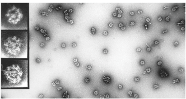
| CRISPER PICTURE Electron micrograph images like this one have revealed the overall shape of the nuclear pore complex (doughnut shapes). Rout?s new method promises to generate a far clearer picture. |
| COURTESY OF MICHAEL ROUT/ROCKEFELLER |
2-D IR Cracks Membrane Protein Structure
|
|
|
HEAD TO TOE Two-dimensional IR spectroscopy confirmed that the peptide AcWLx5, which is a monomer (left) in solution, forms antiparallel  -sheets (right) in lipid membranes. Tryptophan is aqua; the peptide's C terminus is shown in red. -sheets (right) in lipid membranes. Tryptophan is aqua; the peptide's C terminus is shown in red. |
|
COURTESY OF BILL WIMLEY/TULANE |
Determining the structure of a membrane protein within its lipid environment is notoriously difficult. Thus far, only solid-state nuclear magnetic resonance spectroscopy has proven to be up to the task. Paul H. Axelsen reported that he and his colleagues at the University of Pennsylvania have determined the structure of a model membrane protein using a new two-dimensional infrared analog of NMR.
"Our success suggests that this technique will be broadly useful for determining the structures of membrane-embedded proteins," Axelsen told C&EN.
Pioneered by Penn chemistry professor Robin M. Hochstrasser, 2-D IR spectroscopy uses two successive sequences of femtosecond laser pulses to induce molecular vibrations in the protein to be structurally interrogated. The coupling between these vibrations mimics the coupling between magnetic spins in NMR: Vibrations that are within a few angstroms of one another interact and thus give rise to cross-peaks in the 2-D IR spectra. These cross-peaks depend on the protein's secondary and tertiary structures and thus can be used to surmise the protein's 3-D structure.
The 2-D IR technique has been used to probe the structure of small, soluble peptides and proteins. Hoping to extend its utility to membrane proteins, Axelsen chose to study AcWL5, a six-residue acetylated peptide containing five leucines and a tryptophan. The brainchild of biochemist William C. Wimley of Tulane University, New Orleans, this artificial peptide is a monomer in solution, but in lipid bilayers it readily organizes into a higher ordered structure. On the basis of traditional IR spectroscopic experiments, Axelsen and Wimley had previously hypothesized that this membrane-induced structure was an antiparallel -sheet (whereby the peptides stack up next to one another with their tryptophan residues at alternating ends). To prove it, they turned to Hochstrasser's 2-D IR technique.
They found that 2-D IR spectra of membrane-embedded AcWL5 containing a 13C-labeled leucine at either the second or third position reveal a vibrational coupling pattern indicative of an antiparallel -sheet. The coupling patterns also reveal details of the antiparallel -sheet structure that cannot be seen by any other method.
With this demonstration case under their belt, Axelsen and Hochstrasser now hope to use 2-D IR to probe the role of lipid membranes in promoting pathological -sheet structure in protein-misfolding diseases. They are confident that the technique will find broad utility among biophysicists, in part because tweaking the timing of the laser pulses can be used to collect kinetic information. "Two-dimensional IR will soon be used to follow the chemical, structural, and conformational changes of proteins and nucleic acids over time regimes that are not accessible by other methods," Hochstrasser told C&EN.
Protein Clamp Slides Along DNA
|
|
|
KEEP ON MOVIN' Molecular dynamics simulations have shown that, at any one time, a bacterial sliding clamp protein (blue ribbon) traveling along its DNA substrate (center; nucleotide core is green) uses only a handful of the positively charged amino acid side chains (red) in its inner ring to bind to the DNA's phosphate backbone (gold). |
|
COURTESY OF DANIEL BARSKY/LLNL |
In the late 19th century, time-lapse photography by Eadward Muybridge revealed that a trotting horse always has at least one foot on the ground at any given time. Sliding clamp proteins--which play crucial roles in DNA replication and repair--move along the DNA duplex in a similar fashion, according to Daniel Barsky of Lawrence Livermore National Laboratory.
Such proteins clamp like a handcuff around genomic DNA and are thought to slide freely along the DNA like a doughnut on a string, as long as no other proteins are in the way. However, the inner ring of the doughnut-shaped sliding clamp is studded with positively charged amino acid residues that form salt bridges to the negatively charged phosphate backbone of the DNA helix, Barsky noted. "It made us wonder--why doesn't the sliding clamp stay put?"
To address that question, Barsky used the published structure of a sliding clamp involved in bacterial DNA synthesis to perform a 2-nanosecond molecular dynamics simulation of the clamp sliding along a short stretch of DNA. Like the horse in Muybridge's trotting-horse series, the clamp "always touches the DNA--just not with all of its 'feet' at once," Barsky reported. Only a handful of the sliding clamp's positively charged side chains contact the DNA backbone at any one time, allowing the protein to move along its DNA substrate by alternating which set of residues contact the phosphate backbone. Thus the key to sliding "is not fulfilling all of the potential electrostatic contacts at the same time," Barsky told C&EN.
Click image to play video
These moving images are taken from a 2 ns MD simulation of a sliding clamp protein interacting with 12 base pairs of DNA. The simulation contains about 50,000 atoms, including protein, DNA, water and sodium ions. The movies are continuous loops of 100 snapshots, taken every 20 ps. |
|
|
|
|
|
(3.2MB)
|
|
(2.4MB)
|
|
|
| Top view of DNA backbone (tan) and protein (red and blue) and protein residues coming within 3 Angstroms of any DNA atom at that instant. The residue colors indicate positively charged (navy blue), polar (light blue and green), non-polar (white) and negatively charged (red). |
|
Side view of DNA backbone (tan) and protein residues coming within 3 Angstroms of any DNA atom at that instant. The residue colors indicate positively charged (navy blue), polar (light blue and green), non-polar (white) and negatively charged (red). |
COURTESY OF DANIEL BARSKY/LLNL |
|
|
|
|